Stromal-Hematopoietic Interrelationships:
Maximov's Ideas and Modern Models
 
A. Friedenstein    Hämatol. Bluttransf. Vol 32

The Gameleya Institute for Epidemiology and Morphology, Academy of Vet. Med Sciences of USSR. Immunomorphol. Lab., Gameleya Street 18. Moscow D 98, USSR

The idea of stromal-hematopoietic cell interactions was the essential part of Alexander Maximov's theory ofhematopoiesis, which he proposed more than 60 years ago. According to Maximov (see Figs. 1-4), committed hematopoietic precursors descend from the hematopoietic stem cells due to local impacts generated by marrow stroma; this creates the conditions for hematopoietic cell differentiation [1]. Maximov's theory was far ahead of his time, and, though Maximov was highly respected in the scientific community, his concept of local ""differentiation conditions" operative in hematopoiesis was met with particular skepticism. Today, Maximov's idea raises no doubt; in fact, it constitutes the essence of the problem of hematopoietic microenvironment (HME). What provokes discussions in modern hematology is the exact types of stromal cells responsible for HME and the mechanisms of stromal-hematopoietic cell interactions. Maximov assumed that the stromal cells in question were stromal fibroblasts (reticular cells), but for a long time many experimental hematologists denied this. Only recently has it been possible to apply two experimental models for checking the microenvironmental functions of marrow fibroblasts. The first model is the transfer of HME by heterotopic transplantation of marrow cells; the sccond is the establishment of HME in vitro by stromal cell underlayers in Dexter cultures. The Gameleya Institute for Epidcmiology and Morphology, Academy of Vet. Med Sciences of USSR. Immunomorphol. Lab., Gamclcya Street 18. Moscow D 98, USSR Heterotopic transplantation of marrow cells results in the formation of marrow organs covered by a bone capsule [2- 5] .Their hem a topoietic cells are of the recipient origin [6], indicating that engraftment of some category of marrow cells results in the formation of bone and an HME suitable for population by hematopoietic cells and for their proliferation and differentiation. Heterotopic marrow can be retransplanted repeatedly with similar results, provided the recipients are compatible with H-2 antigens of the initial donor, not of the intermediate recipients [7 -8]. This means that HME is transferred by engraftment of the marrow cells which remain unreplaced by the recipient cells. Chromosome typing of clonogenic stromal fibroblasts (CFUf) of the heterotopic marrow confirmed their donor origin [9, 10], and the problem was to check whether stromal fibroblasts were able to transfer HME when grafted heterotopically. The in vitro descendents of CFUf after several passages compose diploid fibroblast culturcs [1113]. Tested by heterotopic transplantation, thcy wcre found to form bone marrow organs, while engraftment of cultured spleen fibroblasts (the descendents of spleen CFUf) produced lymphoid organs [14, 15]. Thus, cultured marrow fibroblasts appear to be able to transfer bone marrow HME. Depending on the origin of marrow fibroblast cultures (the source CFUfbeing from red or yellow marrow), their engraftment transferred not only the general pattern of HME, but also such details as the density of hematopoietic cells in a would-bc marrow [16]. Cultured marrow fibroblasts produce hematopoietic growth factor (M-CSF, G CFS, GM-CFS, BFUf- and mixed-colony-CSF) which can be detected in the culture medium [17- 20]. They regulate proliferation and differentiation of GMCFU: their stimulatory effects were noted when the target marrow contained few spontaneous colonies, the inhibitory effects when large numbers of spontaneous GM-CFU were present [21]. Hematopoietic growth factors are also produced by cloned lines of marrow fibroblasts [22]. However, the direct proof of in vitro microenvironmental competence of marrow fibroblasts was their ability to establish HME in Dexter-type cultures. It has been shown [23] that when used as underlayers, the passaged murine marrow fibroblasts, free from macro phages and endothelial cells, supported hematopoiesis if seeded with stromal cell-depleted marrow suspensions. Thus, cultured marrow fibroblasts transfer HM E, release hematopoietic growth factors in vitro, and are capable of presenting them in a proper way to support hematopoiesis in cultures. This confirms Maximov's hypothesis of the role of marrow fibroblasts in hematopolesIS. The population of marrow fibroblasts is probably a heterogeneous one, and there is no evidence that marrow fibroblasts which produce or present hematopoietic growth factors are the same cells which transfer HME, and vice versa. It may well be that there are several subpopulations of marrow fibroblasts with different microenvironmental functions. At present, fibroblasts including those from nonhematopoietic and hematopoietic organs look much alike, reminiscent of the situation with lymphocytes in Maximov's time. The main and most conclusive sine of fibroblasts (mechanocytes) is interstitial collagen types I and III synthesis, and few markers of their phenotype and genetic diversity have been so far ascertained. The diversity does exist, for instance, between marrow as compared with spleen fibroblasts, which is proved by the results of their heterotopic transplantation. The next question regarding HME seems to be the diversity of marrow fibroblasts including their clonogenic precursor cells. In primary cultures of marrow cell suspensions the CFUf (CFCf) form adherent-cell colonies which are cell clones [24, 25]. The colonies are composed of fibroblasts which synthesize type-1 and -Ill collagen and fibronectin and lack macrophage markers and VIll-factor-associated antigen [26-30]. Morphologically, the colonies are distinctly heterogeneous within each culture. Some are composed of elongated or blanket-like fibroblasts or of a mixture of both; the colonies may include fat cells or have a mineralized intercellular matrix [39]. These differences can hardly be regarded as markers of CFCf, the diversity not beeing stable at passaging and recloning. In situ CFCf are outside the cycle arrested in Go [31]. Marrow fibroblasts possess PDG F receptors [32] and in medium with platelet-poor plasma their proliferation and the CFUf colony formation requires PDGF [33, 34]. It is believed that serum growth factors, which include PDGF, are sufficient for recruitment of CFCf into the cycle and that CFUf colony formation in serum-supplemented medium does not require additional growth stimulation. Yet this is probably not the case. The efficiency of CFUf colony formation (CFEf) drops close to zero in lowdensity marrow cultures if they are depleted of nonadherent cells: 85% of CFCf do not proliferate at all or pass through one to three cell doublings (Fig. 1). On the other hand, the CFEf increases dramatically when such adherent marrow cell cultures are supplemented with irradiated marrow feeder cells or with platelets. This colony-stimulating activity is not replaced by additional PDGF and is expressed only in the serum-rich medium. Being stimulated by platelets each fibroblast precursor present in marrow cell suspensions turns out to be a clonogenic stromal cell (Fig.l). Thus, nonstromal marrow cells which accompany CFCf in marrow cultures


Fig. 1. CFUf colony formation in mice adherent marrow cell cultures. Cultures were initiated by injecting 500.000 mechanically (white colomns) or 50.000 trypzinised (black columns) marrow cells per culture flask (25 cm²). Two hours after cxplantation the nonadherent cells were decanted from all cultures and further cultivation accomplished in aMEM medium plus 20% embryonal calf serum, part of the cultures (G) being additionally suplemented with 10 mio. irradiated (60 Gy) marrow cells. Abscissa: A -E -fibroblast foci, fibroblast colonies and single fibroblasts in feeder non-supplemented cultures. A-single fibroblasts in one day cultures; B-F-10 day cultures. B-single fibroblasts, C-two fibroblasts foci, D-three-eight fibroblasts foci, E-nine-forty nine fibroblasts foci, F- fibroblast colonies composed of 50 and more fibroblasts, sum- sum of B, C, D. E and F per culture. G -fibroblasts colonies in 10 days feedcr-supplemented cultures. Ordinate: mean numbers (M +- m) of single fibroblasts, fibroblast foci and fibroblast colonies for 3- 5 cultures.


(probably megakaryocytes) provide growth-stimulating factors for CFUf colony formation. There are indications that CFCf are sensitive also to other growth-stimulating factors which induce the formation of fibroblast colonies with a different composition of matrix proteins. It has been reported [35] that marrow cells cultured in methylcelluloseclotted plasma with cortisone and PHAstimulated leukocyte-conditioned medium produced fibroblast colonies with collagen type IV and laminin, in addition to collagen types I and III and fibronectin present in CFUf colonies, in liquid cultures with the seruill-supplemented medium. The differences suggest either that there is a diversity of CFCf, which also require different colony-stillulating factors, or that the same CFCf can generate different descendents, depending on the stimulating factors used to induce colony formation. Marrow CFCf diversity was demonstrated with regard to their proliferative and differentiative potencies. Only a small portion (10%) of single CFUf colonies transferred HME when grafted heterotopically, i. e., formed bone marrow organs [36]. At least 30% of CFCf appeared to be highly proliferative cells which provide single-colony-derived fibroblast cultures with 20-30 population doublings. When tested by transplantation of cells in diffusion chambers, 20% of these cultures formed simultaneously bone, cartilage, and reticular-like tissue, 30% formed only bone, and 27% only reticular-like tissue. The number of osteogenic units in late passages of cultured fibroblasts exeeded by far the total numbers of the initially explanted marrow cells, indicating that osteogenic precursors intensively multiplied within cultures [37]. There are reasons to consider CFCf with osteochondrogenic potencies as being osteogenic stem cells [38, 39]. One can assume that some of them are the progenitors of a marrow stromallineage which includes committed osteogenic precursors, mature bone cells, and microenvironmentally competent fibroblasts (reticular cells). The assumption is backed up by the obligatory association


Fig. 2. Type I collagen in 12 day CFuf colony of guinea pig periferal blood leukocytes. Anticollagen antiserum, immunoperoxidase reaction (a). Live culture (b).

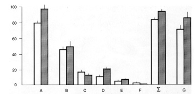
of HME transfer with bone formation, which applies to heterotopic transplantation of both freshly isolated marrow and single-CFUf-derived fibroblast colonies. In the heterotopic marrow the CFUf are of donor origin [9, 10], and it is reasonable to assume that the same applies to the microenvironmentally competent reticular cells. However, the ability of fibroblasts from single CFUf-colonyderived heterotopic bone marrow organs to support hematopoiesis in vitro, and their donor origin (which would be the proof of the above speculation) was not tested up to now. Anyway, the hierarchy of marrow precursors awaits further studies. As far as Maximow's contribution to the problems of HME is concerned, it is impossible to omit his last work, entitled "Cultures of blood leukocytes. From lymphocyte and monocyte to connective tissue." [40]. It describes the formation of fibroblasts in plasma-clot cultures of guinea-pig blood cells. Subsequently, his results were put in question on the grounds of two possible objections, namely that the source of fibroblasts might be fragments of vessel walls which contaminate the blood during sampling, and that the cells in question were not fibroblasts (for references, see [41]). The first objection proved to be invalid when a CFUf colony assay was carried out


Fig. 3. Fibroblasts and collagen fibrils in 16 day CTUf colonies of rabbit periferal blood leukocytes. E. M.

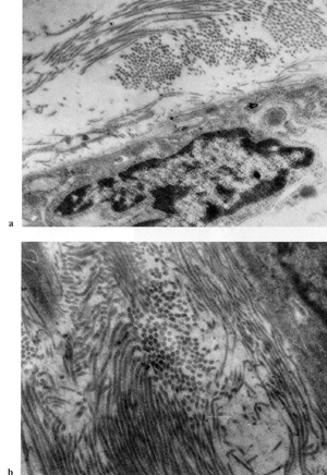
with blood cells. It turned out that the incidence of CFU f colonies in guinea-pig and rabbit leukocyte cultures did not change with 
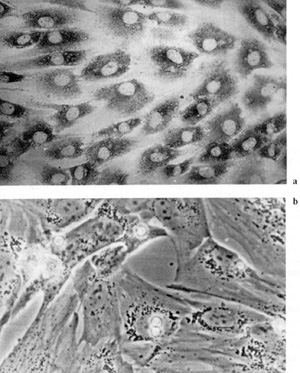
the number of punctures performed for blood sampling [42]. It has also been shown that fibroblasts in blood-derived CFUf colonies synthesize collagen type I [43] and lack VIII-factorassociated antigen and macrophage determinant Mac I [44], which confirms their fibroblast nature (Fig. 2, 3). It remains unknown from where CFUf migrate into blood, where they settle (if they do), and why blood-derived CFUf are not detectable in some mammals, including human beings. The presence of fibroblast precursors in blood discovered by Maximov is related to many unsolved problems of HME, in particular, to the possibility of CFUf repopulation; CFUf circulation in blood does not prove it at all. The story of the circulating fibroblast precursor cells demonstrates once again that not only Maximov's ideas, but also his experimental results are so topical that Professor Alexander Maximov almost remains a participant of presentday research (Fig. 5).


Fig. 4. Porfessor Alexander Maximov


Fig.5.
Maximov in his tissue culture laboratory in the Military Medical Academy in Petersburg (1915)


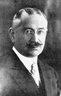
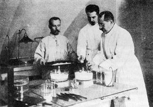
Fig.5a. Preparation of plasma for plasma-clot cultures


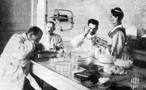
Fig. 5b. Placing tissue fragments in culture medium


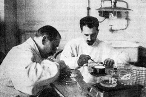
Fig.5c. Kaissug hangrug-drop cultures in
hallowground microscope slides.


References

1. Maximov AA (1906) Über experimentelle Erzeugung von Knochenmarks-Gewebe. Anat Anz 28.24-38
2. Tavassoli M, Crosby WH (1968) Transplantation of marrow to extramedullary sites. Science 161.54-56
3. Tavassoli M, Maniatis A, Bindcr RA, Crosby WH (1971) Studies on marrow histogcncsis. Proc Soc exp BioI Med 138:868-870
4. Tavassoli M, Friedenstcin A (1983) Hemopoictic stromal microenvironmcnt. Am J Hemat 15.195-203
5. Friedenstein AJ, Latzinik NV, Orosheva AO" Oorskaya UF (1982) Marrow microcnvironment transfer by heterotopic transplantation of frcshly isolated and cultured marrow cells in porous sponges. Exp HematoI10:217-227
6. Friedenstein AJ, Petrakova KV, Kuralesova AI, Frolova OF (1968) Heterotopic transplants of bone marrow. Analysis of precursor cells for osteogenic and hemopoietic tissues. Transplantation 6:230-247
7. Friedenstein AJ, Kuralesova A (1971) Osteogenic precursors of bone marrow in radiation chimeras. Transplantation 12: 99-108
8. Friedenstcin A, luria E (1980) Cellular bases of hemopoictic microenvironment. Moscow, Medicine (in Russian)
9. Friedenstein Al et al. (1978) Origin of bone marrow stromal mechanocytes in radiochimeras and in hctcrotopic transplants. Exper Hematol 6: 440-444
10. Friedenstein A (1976) Precursor cells of mechanocytes. Inter Rev Cytol 47: 327 359
11. Friedenstein Al, Chailakhjan RV, lalykina KS (1970) The development of fibroblast colonies in monolayer cultures of guinea-pig bone marrow and spleen cells. Cell Tissue Kinet 3.393 -403
12. Miskarova ED, lalykina KS, Kokorin 1N, Friedenstein AJ (1970) Osteogenic potencies of prolonged diploid cultures of myeloid cells. Bull Exp BioI Med 56.78 81
13. rriedenstein A (1973) Determined and inducible osteogenic precursor cells. Ciba Found Sympos (new series) 11.170-185
14. Friedenstein AJ et al. (1974) Stromal cells responsible for transferring the microenvironment of hemopoietic tissues. Cloning in vitro and retransplantation in vivo. Transplantation 17.331-340
15. Friedenstein A (1980) Stromal mcchanocytes of bone marrow: cloning in vitro and retransplantation in vivo. In. Thierfelder S, Rodt H, Kolb H (eds) Immunology of bone marrow transplantation. Springer-Verlag 19-29
16. Patt HM, Maloney MA, Flannery Ml (1982) Hemopoietic microenvironment transfer by stromal fibroblasts derived from bone marrow varying in cellularity. Exp Hematol 10.738- 742
17. Akasaka Met al. (1987) Production of monoclonal antibody to adult bone marrow preadipocite line (H-IlA). Exp Hemato115:610-618
18. Brockbank K, Van Peer CM (1983) Colony-stimulating activity produced by hemopoietic organ fibroblastoid cells in vitro. Acta Hematol 69.369-476
19. Zucali Jet al. (1986) Interleukin I stimulates fibroblasts to produce granulocyte macrophage colony-stimulating activity and prostoglandin El. J Clin Invest 78.1306-1323
20. lee M, Segal GM, Bagby GC (1987) Interleukin I induces human bone marrowderived fibroblasts to produce multilineage hemopoietic growth factors. Exper Hematol 15.983 -988
21. Brondy Vet al. (1986) Monocytes stimulate fibroblastoid bone marrow stromal cells to produce multilineage hemopoietic growth factors. Blood 68.530-537
22. Grcenbergcr BR, Wilson FD, Woo l (1981) Granulopoetic effects of human bone marrow fibroblastic cells and abnormalities in "granulopoietic microenvironment. Blood 58: 557 -563
23. Brockbank KCM, De long JP, Piersma AH, Voerman JSA (1986) IHemopoiesis on purified bone-marrow-derived reticular fibroblasts in vitro. Exper Hematol 14.386-394
24. latzinik Net al. (1986) The content of stromal colony-forming cells (FCFC) in the mousebone marrow and the clonal nature of FCFC-derived fibroblast colonies. Ontogencsis 1: 27- 35
25. Friedenstein A et al. (1989) On clonality of CFUf-derived marrow stromal colonies (in press)
26. latzinik NV, Sidorovich SY, Tarchanova IA (1980) Studies of surface receptors of the stromal mechanocytes of hemopoietic organs. Immunology 1.26-28
27. Castro-Malaspina H et al. (1980) Characterization of human bone marrow fibroblast colony-forming cells (CFUf) and their progeny. Blood 56:286-301
28. Wilson FD, O'Grady l, Mc Meil GJ, Munn Sl (1974) The formation bone marrow-derived fibroblastic plaques in vitro. Exper Hematol 2.343-349
29. Friedenstein A et al. (1974) Precursors for fibroblasts in different populations of hemopoietic cells as detected by the in vitro colony assay method. Exper Hematol 2.83-92
30. Friedenstein Al, Gorskaya UF, Kulagina NN (1976) ribroblast precursors in normal and irradiatcd mouse hemopoictic organs. Exper Hematol 4.267 -274
31. Keilis-Borok IV, latzinik NV, Epichina SY, Friedenstein AJ (1971) Dynamics of the formation of fibroblast colonies in monolayer cultures of bone marrow, according to ³H-thymidine incorporation expcrimcnts. Cytologia 13.1402-1409
32. Bowen-Pope DF, Seifert RA, Ross R (1985) The platelet-derived growth factor receptor. In. Boyton l, leffert H (eds) Control of animal cell proliferation, Acad Press 1.281-312
33. McIntyre AP, Bjornson BH (1986) Human bone marrow stromal cell colonies. response to hydrocortisone and dependence of platelet-derived growth factor. Exper Hematol 14: 833- 839
34. Wang OR, wolf NS (1987) The effect of several growth factors on the in vitro growth of bone marrow stromal cells. Exper Hemato115.610 35. Lim Bet al. ( 1986) Charactcrization of reticulofibroblastoid colonies (CFU-RF) derived from bone marrow and long-term marrow culturc monolayers. J Cellul PhysiolI27.45-54
36. Chailakyan RK, Gerasimov YF, Friedenstein AJ (1984) Content of osteogenic precursor cells in the bone marrow and their prolifcration in cultures. Bull Exp BioI Med 11.605-608
37. Gerasimov Y, Friedenstein AJ, Chajlakjan RK, Shiskova VV (1986) Differential potentiality of clonal strains ofbone marrow fibroblasts. Bull Exp BioI Med 6.717 -719
38. Friedenstein AJ, Chajlachyan RK, Gerasimov YF (1987) Bone marrow osteogenic stem cells in vitro cultivation and transplantation in diffusion chambers. Cell Tissue Kinet 20: 263- 272
39. Owen ME, Friedenstein AJ (1988) Stromal stem cells: marrow derived osteogenic precursors. In: Evered D, Harnett S. (eds) Ccllular and molecular biology of vertebrate hard tissues. Ciba Found Symp 136.42-52
40. Maximov A (1928) Cultures of blood leucocytes. From leucocyte and monocyte to connective tissue. Arch exp Zellforsch 5.169-178
41. Jacoby F (1965) Macrophages. In: Willmer E (ed) Cells and tissues in culture, Acad Press 1.1 -93
42. Luria EA, Panasyuk AF, Friedenstein AJ (1971) Fibroblast colony formation from monolayer cultures of blood cells. Transfusion 11.345-349 43. Luria E et al. (1989) Colony forming fibroblast precursors in circulating blood. Bull Exp Biol Med (in press)
44. Piersma AH, Ploemacher RE, Brockbank KG (1985) Migration of fibroblastoid stromal cells in murine blood. Cell Tissue Kinet 18:589-595