|
* This work was supported by NIH Grant Nos. CA 28461. CA 29609. CA 14216, and A1 07019 Contrasuppression is an immunoregulatory T -cell activity that protects Lyt I+. 2- T helper cell activity from suppression. This activity involves both an "induction" (afferent) phase, which requires the activation of an Lyt 1+, 2- effector T cell by cells in the contrasuppressor circuit [6], and an "effector" (efferent) phase, in which the effector cells or cell-free products secreted by these cells render T H cells resistant to suppression [4]. Recently we discovered an activity in antisera raised against methylcholanthrene-induced sarcomas from Balb/c mice, which blocks T -cell regulatory activity [3]. These antisera block the afferent as well as the efferent phase of suppression to SRBC in vitro, but only in animals which express the same Igh gene polymorphism as Balb/c (lgha). We therefore tested whether these antisera could block the afferent and efferent phases of contrasuppression. and whether this activity had any effect on the growth of tumors in those mice.
The chemically induced sarcomas, and the antisera against them, were prepared according to procedures described by DeLeo et al. [ 1. 2] .Suppressor T cells were prepared according to the method of J aneway [5]. Contrasuppressor T cells were prepared according to the method of Green [4]. Contrasuppressor factor (T csF) is a cell-ffee supernatant collected from in vitro generated T cs cells. Generation of primary antiSRBC cultures and blocking assays with antisera has been described [3]. Assays for metastasis were performed by injecting 10 high5 or 5 X 10 high4 Meth A cells into the right footpad of test animals. After 3-4 weeks, lymph nodes were removed and weighed and examined histologically for evidence of tumor cell growth. Animals positive for metastasis were those which showed tumor cell growth in the popliteal lymph nodes of the left leg, as well as both axilary lymph nodes.
Antisera effective in blocking the afferent but not the efferent phase of suppression were tested for their abilitiy to block the afferent and efferent phases of con trasuppression (Table I). Antisera raised in syngeneic Balb/c mice against Meth A (or other MC-induced tumors. data not shown) were ineffective in blocking the activity of either the T csF , which represents the efferent phase of contrasuppression. or the T cs cells. which represents the afferent phase of contrasuppression. However. antisera raised in semisyngeneic CB6Fl or 19h congenic C.B20 mice effectively blocked the activity of the Balb/c T cs cells but not the T csF. Likewise. these antisera were very effective in blocking afferent Tcs activity in CB6F 1 mice, while they were ineffective in blocking T cs activity in Lgh disparate mice, reiterating the earlier finding on the nature of Table I. Antisera to Meth A raised in
Ighb+mice block contrasuppression 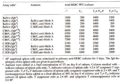
Table 2. Tumors passed in CB6Fl but not
Balb/c absorb blocking activity 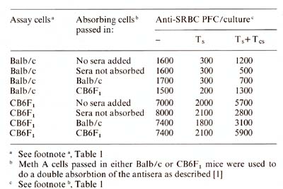
An additional activity, the blocking of contrasuppression, has been found in antisera against MC-induced tumors. Experiments with F 1 and Igh congenic mice indicate that effective antisera can only be generated in mice containing Igh disparate genes, while activity is only directed against cells expressing the Igh a gene locus. This brings up the apparent dichotomy that F 1 mice generate autoantibody to their own Ighlinked gene products. However, tumors passaged in F 1 animals express the relevant antigen in a much higher surface density than does the parental strain. This ""adaptive differentiation " process may explain the difference in tumorgenicity between F 1 and Balb/c mice, as measured by metastasis. The intriguing possibility exists that F 1 mice produce autoantibodies that block the generation of their own contrasuppressor cells, and that these contra suppressor cells are important in controlling tumor metastasis. It also suggests that while many tumor cells escape immune destruction by generating suppressor T cells to depress immune responses, malignant cells may also escape by ""encouraging" immunity, e.g., generating antigens which mimic normal cellular interaction structures and thereby blocking important cellular communication mechanisms needed to generate effective antitumor immunity.
1. DeLeo et al. (1977) J Exp Med 146: 720 2. DeLeo et al. (1979) Proc Natl Acad Sci USA 76:2420 3. Flood et al. (to be published) Proc Natl Acad Sci USA 4. Green et al.(1981)EurJlmmunol ll:973 5. Janeway et al. (1975) Nature 253:544 6. Yamauchi et al. (1981) J Exp Med 153: 1547 |