|
Hans Eidig Lecture
Wilsede, June 23, 8pm 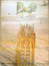
Introduction to Hans Eidig and Robert C. Gallo An introduction to Bob Gallo, as a person and a scientist, would
certainly be superflous. I would therefore simply like to draw a
parallel to the life of the lectures eponym, Hans Eidig. The name
of this special lecture reminds us not only of particular historical
circumstanCes but, more importantly, of this man's life. Hans Eidig
was born in 1804 in the village of Klein Klecken. It was originally
intended that he should become a forest ranger, as was his father.
However the yearning for freedom and independence, which was to
innuence his entire life led him to become a poecher rather than
a forester. He was very good at this, and there was a great demand
for his services. But his desire for freedom and independence was
too strong for the restraints of his way of life, not afraid of
this risking his life for others, he became the Robin Hood of the
Lüneburger Heide. He had good friends and advisors as well as many
enemies. In 1835 he agreed to the king's offer to accept the cash
reward on his head and to emigrate to America. He arrived in New
York with his girlfriend, dreaming of freedom and independence.
Thereafter we largely lose track of his life in the new continent.
However, those of us from this region believe that he traveled to
the West Coast of the United States where he discovered gold. Although
I do not know whether this legend is true, it remains very much
alive here, and today's inhabitants of this village remember him
as a local hero who always tried to protect our ancestors from humiliation
at the hands of their feudal masters. In the New World Hans Eidig
gained the freedom and independence that he had always sought. Against
the background of these words honoring the memory of Hans Eidig,
I should like now to present Bob Gallo, who will explain the origin
of human leukemia. Personal Reflections on the Origin of Human Leukemia Before I finish this lecture everyone else is going to be dreaming of freedom and independence. Rolf Neth wishes to link me with Hans Eidig. I may indeed have three features in common with this fellow, this Robin Hood as you call him. These are: very good friends, at least a few good enemies, and some foolishness - foolishness to give a lecture at this hour about such a topic. First, I have been asked to give my reflections of the Wilsede meetings since their origin some 15 years ago and, then, on the virus "hot spot" theory, based on an article that I wrote for the first Wilsede meeting. That article was an attempt to counter the overused and overstated virogene/ oncogene theory as it was originally proposed. We have, however, progressed far past that stage. About the beginning of the meetings. In 1970 or 1971 I first met Rolf Neth. We walked along the Hamburg harbor and later we came to this forest he loves so much. He catalyzed (almost immediately) a similar affection from me. I told him that this would be a great place for meetings, regular meetings to promote friendly informal discussions among friends and adding new people as time went by. By 1970 there was much activity developing in basic cancer research and all its subtopics. I felt those dealing with blood cell biology and the leukemias and lymphomas would make the greatest advances. First, because we could get our hands on such cells; secondly, because so many animal models were available, and, thirdly because there seemed to be, in general, a high caliber of people involved in research in that area of cancer. I also believed that it was time to push human studies to the point that they would be scientifically acceptable. So I though that if we had small meetings with a percentage of people always returning and then in time, adding people who get interested and who had particularly interesting information from diverse disciplines but linked by some interest in blood cells, lymphocytes and/or their malignant transformation, we could enhance development of the field. The atmosphere of this marvelous forest was to augment the interactions. In time we hoped research in human disease would no longer be frowned on by the intellectual lights. To signal this feeling, we incorporated "human" in the meeting titles. Av Mitchison, Peter Duesberg, Rolf and Malcolm Moore and Mel Greaves gave the spark and life to the early meetings, as, of course, did the great Fred Stohlman at the first. Now forced to think back on all those years, I don't know if there's disappointment or elation in the progress the field has made nor whether we achieved our objective. The biggest disappointment I had is that at the time those meetings were organized, everyone was talking about multiple causes or primary agents or whatever we wanted to call the true causes ofleukemia and lymphomas, but we all thought there would be one common mechanism at least one common, final biochemical mechanism. That doesn't turn out to be the case. At the beginning of this century, the idea of leukemia most people pictured was that of a very wild disorder of cell proliferation. Later, with advances in the understanding of pernicious anemia (in which erythroblasts resemble malignant cells), the induction of abnormal cells to differentiate into normal cells using a simple vitamin replacement, vitamin B,,, came the opposite polar extreme idea that leukemia was simply a nutritional disease. One specific factor might take the whole thing away. Certainly by the time of these meetings, a position very much in between was already in hand. Around 1950, as I remember the literature, a group in northern Italy, led by G. Astaldi and F. Gavosto and their co-workers, published a series of papers on thymidine radioautographs of labelled cells, concluding that leukemic cells do not wildly proliferate, but, in fact generally, have longer generation periods than do normal cells. By the first meeting people had already accepted the idea that leukemia was a kind of a block or a frozen state of differentiation, maybe partly reversible, maybe not. During that period of time, just before the meetings began, the concept of monoclonality had predominantly came from people who were doing chromosome studies, and made unambiguous by the studies of Phil Fialkow and his colleagues who used G-o-PD variants as X-linked enzyme markers to demonstrate monoclonality of CML. These studies were described at these meetings. So we had the concept of clonality of a partial or complete block in differentiation and presumed that a common molecular mechanism might account for all. At that time also most people thought stem cells were the only targets of a putative leukemogenic agent. This was championed, but it is not to be the case. Today we would say any cell capable of continued proliferation, even if committed, can be a target of a carcinogen or a leukemogen. The first meeting began about 4 or 5 years after the discoveries by Leo Sachs and his co-workers and by Donald Metcalf and Bradley which led to a reproducible system of growing cultured cells in colonies and growth factors for leukocytes. The first blood cell growth factor had, in fact, already been discovered many years before by Allan Erslev, my first scientific mentor when I was a medical student. This was by the growth factor erythropoietin for red cells and was partly defined at the time of the first Wilsede. However, the field of leukocyte biology really progressed by the clonal assay systems. By the first meeting, we had the idea (at least for myeloid leukemic cells) that the block in differentiation could be overcome, at least in part. Leo Sachs talked here about the uncoupling of growth and differentiation and proposed that the molecules for these are in general separable. Both of these pioneering groups and their colleagues, especially Malcolm Moore, developed the concept that the "blocked" differentiation was not absolute and that some leukemic cells could at least be partially differentiated. Dunng this penod, people also began to get a handle, not only on CSF and the related CSF molecules, but on other growth factors and their receptors. Not only the proteins, but eventually also the genes for some. The earliest of these was interleukin 2 (IL-2) and its receptor. At the same time, so-called protooncogenes became defined. We should remember how those terms came about, and what an oncogene really meant, and what is has become to mean today. Perhaps the word today is used a bit too loosely. These genes were defined as ones capable of transforming a XXXIV primary cell. They were first genetically and molecularly defined by groups in California, notably our own, Peter Duesberg, Peter Voigt and, also, by S. Hanafusa and Michael Bishop. Thus, the first transforming genes were being defined at the time of our earliest meeting and became a frequent topic of discussion - as they continue to be. Dominic Stehelin working with Bishop and independently some people at NIH, Edward Scolnik, Peter Fishinger, and Ray Cilden obtained data that the oncogenes of viruses were derived from the genes of normal cells, but had evolved away from those genes of normal cells. With those defined oncogenes in hand from animal viruses we could capture analogous genes from cells for the first time. Probes were now available to go into normal cellular DNA to "fish out" those homologs of retroviral oncogenes. The genes in normal cells were then called protooncogenes. We could explore the presence of these genes, what state they were in, i.e., amplified - nonamplified, rearranged or not, expressed or not and whether their coding was related to that of known genes for proteins important to growth, e.g., growth factors and their receptors. For the first time, genes and gene products suspected to be important for cell growth and/or differentiation of normal and leukemic cells could be compared. In the past 4 or 5 years or so several results have indicated that these previously independent fields will merge, because certain protooncogenes have indeed turned out to be genes for growth factors or receptors. The c-sis gene for the platelet-derived growth factor, the ErbB gene as a truncated EGF receptor, and recently thefos gene as the macrophage colony stimulating factor receptor are cases in point. At the earliest meetings, I remember the good debates between Jane Rowley and Henry Kaplan on another area of leukemia/lymphoma research, relating to the relevance of chromosomal changes and whether these constitute anything other than secondary effects. As I remember it, Henry Kaplan was always asking for direct evidence that specific chromosomal changes were important for the initiation and/or progression of leukemias and lymphomas. In 1971-1973 the only clear-cut example of this available to us was that of chronic myelogenous leukemia and the Philadelphia chromosome. At about that time the concept that a balance of genes was important to control cell growth was also being widely discussed. Studies from childhood cancers with consistent chromosomal deletion and those of Fritz Anders on fish melanoma provided us with concepts of regulatory genes which control other genes that are more directly involved in cell growth. The chromosomal changes that occur in human leukemia still raise some questions. The first problem as I see it, is why are there specific "hot spots" in human chromosomes that undergo change? In a recent review Mittelman has shown the accumulation of chromosomal breaks in a variety of human leukemias; it is evident that there are sites that must be especially fragile. Another interesting question that I rarely hear discussed is why adult tumors show progressive subclone heterogeneity? This is less common in childhood tumors, but in adult cancer we always talk about progression of the cancer and heterogeneity of subclones as though this is natural and should occur. But can anyone really explain them? It's usually said that this is due to an alteration in genetic regulation in addition to the primary chromosomal abnormality; however, this would imply that an alteration in regulators regularly occurs. Does anything specifically cause the chromosomal change? There are at present no specific molecular mechanisms or inciting agents that have been proven to cause any of the important chromosomal changes. I believe that everyone would accept the idea that specific chromosomal changes are com- XXXV mon and are probably very important to the pathogenesis, if not to the origin, of many human leukemias and lymphomas. In my own view, three genes deserve special discussion, for research into them seems most exciting. These are the c-abl gene in chronic myelogenous leukemia, the c-myc gene in Burkitt's lymphoma, and perhaps the chromosome 5 deletions in myeloid leukemia. The cluster of CSF genes and the receptors for CSF in one region of chromosome 5 suggests that any change in this region is likely to be important for leukemia. Obviously, when there is a specific chromosomal change occurring regularly in a certain leukemia, one would expect it to be important. When there are regions coincident with the location of genes important for the growth and differentiation of a particular cell type, we logically attach special attention to it. The first consistently observed chromosomal abnormality, the Philadelphia chromosome, was discovered by Hungerford and Nowell in the early 1960s. This was first thought to be chromosome 21 deletion but was later shown to involve a translocation between chromosome 9 and chromosome 22. When we had a handle on the gene, several laboratories, including my own, were able to localize the c-abl gene to a region in chromosome 9 where the break occurs. We now know that this gene is translocated to chromosome 22, adjacent to a gene given the abbreviation BCR. Shortly thereafter Canaani at the Weizmann Institute in Israel discovered an abnormal c-abl messenger RNA in chronic myelogenous leukemia. This seems to be an area worthy of major investment. The study of the nature of this gene product might determine the function of the normal c-abl, explain why blast crisis develops, and eventually obtain evidence as to whether leukemia transformation and blast cnsis are directly related to the abnormal c-abl product. An increase in number ofchromosome 22 is common in CML blast crisis. Is this associated with an increased dosage of the c-abl messenger RNA? Probably so, but still, like in almost the entire field, we're left completely wanting for an explanation at the biochemical (mechanism) level of what the molecular genetics has defined. Another approach that has been principally explored and pioneered by Carlo Croce and discussed in detail at this year's meeting, has been to examine cellular nucleotide sequences near chromosomal breaks consistently where there are no known oncogenes and, hence, no easily available or known molecular probes. This considerable endeavor is done by sequencing all around the chromosomal break point and determining which, if any, of these sequences near the chromosomal breaks are abnormally expressed. These can then be molecularly cloned and studied in detail. In other words, these sequences are not homologues of any known oncogene, nor do we have information that they code any growth factor or growth factor receptor. They are identified and worked with solely because they are sequences or genes located near a known consistent chromosomal break. This approach is logical and ver~ likely important way to proceed. The problems are knowing what the gene product does and proving that it is important for the development of the tumor. The last sequence I wish to discuss is that of the cellular homologue of the myc gene. We reported at one of the Wilsede meetings an analysis of the human c-myc gene. This was at a time when the abnormal chromosomes in Burkitt lymphoma were well known (generally 8 and 14). At these meetings George Klein and his associates showed that this phenomenon involves an 8:14 translocation. Independently, Carlo Croce showed that this same region of chromosome 14 involves the heavy chain loci of immunoglobulin XXXVI genes. Subsequently, Riccardo Dalla Favera, a postdoctoral, cloned and mapped the human c-myc gene for the first time. Then together, with Flossie Wong-Staal, he formed a collaboration with Carlo Croce and then demonstrated the location of c-myc at the distal end of the long arm of chromosome 8. In later studies we reported that c-myc was translocated from chromosome 8 to chromosome 14 in each Burkitt lymphoma. The results were also presented and discussed in detail at these meetings. Now, of course, it is known that translocations also involve chromosome 8 and chromosomes 2 and 22. P. Leder, C. Adams, S. Cory, G. Klein, P. Marcu, T. Rabbits and particularly C. Croce and their colleagues have made major contributions to our understanding of the details of the various translocations and their significance. This give me an opening to discuss a few aspects of the epidemiology of leukemias and lymphomas. Burkitt lymphoma (BL) seems to be a classical example of the multistage, multifactoral, multigenetic series of events said to be prerequisites for the development of a malignancy. We would all probably agree that the Epstein-Barr virus (EBV) plays a role in this malignancy, but the generally precise geographic limitation of BL means that its development requires at least one additional environmental factor, and holoendemic malaria is believed to be one such factor. The malarial organism apparently not only provides chronic antigenic stimulation but may also alter T-cell function in such a way that cytotropic T cells do not properly control EBV. So there appears to be three key events: the presence of EBV, the presence of chronic antigen stimulation, and possibly a change in T-cell function. Furthermore, the available evidence argues that during B-cell gene rearrangement a chance translocation of the myc gene occurs, and that this leads to one step in the tumor origin. The probability of this event is presumably increased by the chronic antigenic stimulation change in T-cell function and the excess replication of EBV. The last twist comes from new data that seems to argue that even this activation of the c-myc gene is not sufficient, and that there must be still another event. Susan Cory will present evidence at this Wilsede meeting that many cells of transgenic mice have translocated activated c-myc genes but a tumor arises from only one such cell. Thus, this disease demonstrates multifactoral, multistage, probably multiple genetic events in a cancer. But can this serve as a general model? Should we think about all leukemias, lymphomas, and cancers as multifactoral, multistage and multigenetic? There are some things that bother me about this conclusion. For example, Kaposi's sarcoma occurs in a high percentage of homosexuals with AIDS. Does that mean that these people run around with multiple genetic events already in their endothelial cells just waiting for a T4 cell depression? I believe it much more likely that this is due to the requisite genetic changes occurring with one or, at most, two events; these changes probably include the addition of new genetic change from infection with a virus yet to be discovered. Another apparent exception involves cancers occurring in young girls whose mothers had received estrogen during pregnancy, and who at age 13 or 14 may develop vaginal cancers. Can we see this as multistage, multifactoral, multigenetic events? Also, what about the T-cell leukemias associated with HTLV-I? Because of the considerable time between infection and the leukemia, it is clear that there is more than one stage and probably more than one genetic event, but I know of no evidence that other exogenous factors are required. All current data argues that the virus and the virus alone is sufficient. Regarding most other leukemias and lymphomas, looking for inciting agents or "true primary causes" has been difficult and not very productive to date. It would be disappointing if it turns out there are no initiating agents in most of the other leukemias or lymphomas, because this would mean most are chance events and mean we have nothing to do other than the laborious protein chemistry and metabolism studies like Boyd Hardesty and his colleagues have been doing and reporting at these meetings. Some epidemiological studies demonstrated the importance of radiation, e.g., the Atomic Bomb in Japan and occupational sources of radiation in mostly myeloid leukemias. Chloramphenicol and benzene exposure have also been reported to be associated with an increased incidence of some leukemias. How can we think of chloramphenicol and benzene in causing leukemia? Perhaps they alter programs of gene expression and allow some cell clones to emerge that have been genetically altered by other agents. Although these few chemicals and some forms of radiation are linked to an increased incidence of leukemia, it is, of course, only a very small fraction. For the vast majority of such cases no environmental factor(s) have yet been found. Retroviruses in animals and also in humans (at least since 1979) have been frequently reported and passionately discussed at the Wilsede meetings from the outset; these have been the special interest of many investigators here, including myself. This interest came from the numerous and diverse leukemia-causing animal retroviruses that were already available when these meetings began. Many of us believed (and still do) that these animal models provide powerful tools for an understanding of the molecular and cellular pathogenesis of leukemias/lymphomas and of the genes involved. Some of us also believed that through studies of them we might also learn how to find similar viruses in humans if they existed. When we recall the first meetings at the beginning of the 1970s, the retroviruses that were then discussed most thoroughly and most commonly (and those best supported financially) were the endogenous retroviruses. The genes for these viruses exist in the germ line and are present in multiple copies; and most, if not all, vertebrates and even some other species contain these genetic elements. Sometimes these are capable of giving rise to a whole virus particle. These were the major early focus of studies in leukemogenesis. For example, Henry Kaplan's first studies of radiation leukemogenesis suggested that radiation induced the expression of an endogenous virus which is critical to the development of the leukemia in this case in mice. Huebner and Todaro's onginal theory maintained that all cancer is due to the activation of these endogenous viruses, i.e., to oncogenesis, and was invariable due to expression of endogenous viral genes. This idea in its original and literal form has now been discarded. Ironically, the only retroviruses known to be involved in cancer in humans or in animals (except for a few very inbred mouse strains) are infectious (exogenous) retroviruses. We began to focus on exogenous retroviruses in animals and humans in 1970 after the discovery of reverse transcriptase by Howard Temin and David Baltimore. A major reason for me to focus on exogenous viruses was the innuence of people like Arsene Burny and his studies of bovine leukemia, William Jarrett on feline leukemia, and later those of Max Essex. These researchers, doing veterinary biology-virology, argued that naturally occurring leukemias and lymphomas in animals are apparently often due to exogenous retroviruses. At that time the concept of any cancer being infectious was thought to be naive at the very best. Subsequently, we learned that investigators (again, usually veterinary biologists) working in the avian systems had shown even earlier that an exogenous infecting retrovirus, known as avian leukosis virus, was a major cause of leukemia and lymphoma in chickens. Moreover, a closer examination of the murine leukemia virus literature suggested that mouse viruses may infect the developing offspring in utero or shortly after birth and enhance the probability ofleukemia. Although a vertical transmission, this was, according to Gross, still an infection and not the simple gene transmission of unaltered endogenous retroviruses. In the 1970s we obtained our first primate model. A Japanese-American, T. Kowakami, discovered the gibbon ape leukemia virus (GaLV); he showed that this retrovirus caused chronic myeloid leukemia in gibbons and that a variant of it causes T-cell acute lymphocytic leukemia. My coworkers and I isolated still another major variant of GaLV and we had the opportunity to study gibbon leukemic animals in detail, demonstrating the exogenous nature of GaLV and determining the presence of provirus in the tumor, and analyzing the GaLV genome. Dr. Flossie Wong-Staal in our group also showed that another newly isolated simian retrovirus, known as simian sarcoma virus (SSV), or woolly monkey virus, had viral sequences essentially identical to GaLV but contained additional sequences specifically homologous to cell sequences of the normal (uninfected) woolly monkey DNA. Moreover, Wong-Staal et al. and others showed that GaLV had homologous sequences in the DNA of normal mice, particularly in that of some Asian mice. Erom all these results we concluded that GaLV was an old infection of gibbons (many gibbons are infected in the wild), and that it entered these animals by way of an interspecies transmission of one of the Asian mouse endogenous retroviruses, perhaps by some intermediary vector, we also concluded that the woolly monkey virus (SSV) was derived by an interspecies transmission of GaLV from a pet gibbon to a pet woolly monkey housed in the same cage. (This history was verified by the owner of these animals and by studies which showed that woolly monkeys in the wild are not infected.) This resulted in a recombination of the viral sequences with cell sequences of the woolly monkey. These cellular sequences were later shown to be genetic sequences ofplatelet-derived growth factor (PDGF) as discussed by Aaronson and coworkers, Westermark and coworkers, and others. These results influenced our thinking and the direction of our research. From this point, we considered the notion that a human retrovirus may have little or no homology to human DNA. With rare exception, this was a concept hitherto not even considered by the field. Animal retroviruses, including the disease-causing exogenous FeLV and avian leukosis virus (ALV), although clearly not endogenous, genetically transmitted elements clearly were substantially homologous to DNA sequences of the infected host cell in cat and chicken respectively. The results with GaLV showed little or no uninfected gibbon apes or woolly monkeys. All of these concepts and results have been detailed at previous Wilsede meetings. While we were still considering the possibility (in our view, probability) that human retroviruses would be found, we were, nevertheless, concerned that in these animal models (FeLV, ALV, MuLV, and GaLV) of retrovirus leukemias, virus was so readily found as to require no special techniques or efforts. In fact, viremia preceded leukemia, and it was frequently argued that extensive viremia is a prerequisite for viral leukemia. These facts had been presented as strong arguments against the existence ofa human retrovirus and against the need for any special sensitive techniques to find them. Moreover, it was during the period of the first few Wilsede meetings that a few candidate human retroviruses turned out to be false leads, such as the RD114 virus and the virus called ESP-1, which were shown to be a new endogenous feline virus and mouse leukemia virus respectively. Both were contaminants of human cells. Two model systems developed in work with animals - FeLV and BLV helped sustain the thinking that human retroviruses probably do exist. Although FeLV was known to replicate extensively in most cat leukemias/lymphomas (see above), many of these cases were virus negative, as William Hardy and Max Essex emphasized at early Wilsede meetings. The epidemiology of these cats strongly implied that FeLV was involved. In addition, although virus could not be found in the tumor cells, it was often found at low levels in a few cells of the bone marrow. Therefore, Veffa Franchini, S. Josephs, R. Koshy and Flossie Wong-Staal in our group, pursued molecular biological studies of these tumors in collaboration with Essex and Hardy. We suspected that defective (partial) proviruses might explain the phenomenon. However, we were not able to prove this, and these interesting findings - as detailed at previous meetings - still lack an explanation. Presumably FeLV is involved in these leukemias by an indirect mechanism and not by a provirus integration into the cell destined to be the tumor. The second example of an animal model system which became extremely important for human retroviruses is that of the bovine leukemia virus (BLV). This too was discovered at the beginning of the 1970's when Wilsede meetings were just getting underway. Since its discovery in Iowa by Van der Maartin and coworkers, much of the work on BLV biology was studied by Ferver et al. in Philadelphia and by Arsene Burny and his group in Brussels. It was also Burny et al. who carried out virtually all the BLV molecular biology. At the very first Wilsede meetings Arsene emphasized the minimal replication of BLV, the lack of viremia in infected animals, and the rare expression of virus in the tumor despite the presence of an integrated provirus. This was, of course, precisely the situation with HTLV-I and -II. Ironically, several features found for HTLV-I were later then found applicable to BLV. There may be a lesson for us in this brief history: if our interest is a human disease, we should not allow ourselves to be trapped into focusing upon only one animal model but rather look more broadly at cell models. During the mid 1970's and using the available monkey and ape viruses to make immunological and molecular probes, numerous groups including ours, reported finding virus-related molecules in some human cells, especially leukemias (our laboratory, Fersten group and Peter Bentvelyen and colleagues). The viruses were subsequently shown to be extremely closely related to the simian sarcoma virus (SSV) and GaLV or identical to them. Again, these studies were detailed in Wilsede meetings. Since there has been no further progress with these categories ofvirus, we must at least tentatively believe they were laboratory contaminants. Nonetheless, there are many indications that lead me to think that it will be interesting in the future to reevaluate the question of retroviruses related to GaLV and SSV in humans. In the remaining part of this presentation I will summarize our information on the known existing human retroviruses and highlight a few of the events that eventually led to their discovery. When we began a search for human retroviruses beginning and reported at this first meeting it was in parallel with the late Sol Spiegelman and his colleagues, such as Arsene Burny and Rüdiger Hehlmann. Three approaches were used. As mentioned above, both Spiegelman's laboratory and ours exploited reverse transcriptase (RT) as a possible sensitive assay for discovering these viruses. Perhaps we could detect low levels of virus. Between 1970- 1975 the methods were made more sensitive and specific. The latter was necessary in order to distinguish viral RT from normal cellular DNA polymerases. These techniques, including the development and use of new synthetic homopolymeric template primers has been detailed in several reports. We were able intermittently to detect an enzyme which looked just like the viral RT, and we believed it might be a marker for an exogenous infecting retrovirus. Spiegelman's group also used another approach that gave tantalizing results that most people couldn't quite accept, but I know of no one who has taken the trouble to re-evaluate these experiments. This approach made use of cDNA copies of messenger RNA transcripts from leukemic cells and showed some to be leukemia-specific, i.e., extra to the leukemic cells not present in normal cellular DNA. In at least a few cases Spiegelman and his colleagues could argue that these sequences were partly homologous to sequences present in some animal leukemia viruses. Thus, these experiments suggested that human leukemic cells contained added and probably virus-denved sequences. No one has yet pursued these studies further. The key criticism has always been that the amount of difference between the hybridization to leukemic cell DNA versus normal cell DNA was very extremely slight. The second approach mentioned above, and one that we were lucky to take, was based on our attempts to define various growth factors for human blood cells, partly to help in our pursuit of a human retrovirus and partly because of S>ur interests in blood cell biology. Initially we were looking specifically for a granulopoietic factor to grow granulocytic precursors. Our view for using a growth factor to help find virus was the belief that if a virus was present in low amounts, we could amplify it in this way, and if a viral gene was not expressed, we might induce its expression by growing the cells for a long period of time. Thus, we were looking in any event for a growth factor that would grow granulopoietic cells, not, as Metcalf and Sachs had done, for colonies, but in mass amounts in liquid suspension. We had some temporary success in this. In these efforts we included conditional media from PHA-stimulated human lymphocytes, because in 1971-1972 we and others had discovered that stimulated lymphocytes released growth factors for some cell types. It was while looking for the granulocytic growth factor that we discovered IL2, or T-cell growth factor, cntical to our work. This discovery was made by Frank Ruscetti, the late Alan Wu and especially Doris Morgan. We also learned that activated but not "resting" T cells developed receptors for this growth factor. Peter Nowell had shown in the 1960s that after PHA lymphocytes live and grow for approximately two weeks, they tend to be lost and die out. We used the media, fractionated it, and added fractions back to the same stimulated cells. As long as we kept adding growth factor, the normal T cells continued to grow for considerable periods of time. We then approached studies ofleukemic T cells; with one type ofleukemia, the T cells grew as soon as we put them in culture and added IL-2. We did not need to activate them. They grew directly and they gave rise to the viruses that we have called HTLV-I. The first human being from whom a retrovirus was isolated lived near Mobile, Alabama, in the south east of the United States. This man had no medical, personal, or social history when he developed a very aggressive Tcell malignancy. This was late 1978 when I called Arsene Burny for BLV reagents which, as Arsene likes to remind me, was on Christmas Eve. By that time we knew that the virus from this man behaved somewhat similar to bovine leukemia virus: it replicated poorly, seemed to cross-react slightly with BLV, and morphologically looked more like BLV than like other retroviruses. We thought, however, that we must rule out a bovine leukemia vin~s contaminant in the calf serum used in culturing human T cells. With the reagents to BLV provided by Arsene Burny this was done and soon we were able to charactenze HTLV-I and to report on it at the beginning of 1979. We then published a series of papers on this in 1980 and early 1981. The clinicians called the malignancy an aggressive variant of mycosis fungoides (MF). Also known as cutaneous T-cell lymphoma, this is a T4 malignancy with skin manifestations, lymphoma cells infiltrating the skin, and usually a prolonged course. We obtained a second isolate within a few months; this was from a young black woman in New York City who had come there from the Caribbean Islands. As in the case of the first patient, her illness resembled MF. It now seems, however, that MF may represent more than one disease (all with similar clinical manifestations), and that several forms of T-cell malignancies that are not MF may mimic that disease. By early 1981 the several sporadic cases which we had studied showed the presence of HTLV-I, all involved T4 cells, and all had an acute clinical course, often with hypercalcemia. Tom Waldmann of NCI told me at that time of studies which had been conducted in Japan and published by Takatsuki, Yodoi, and Uchiyama. In 1977, they described a disease cases ofwhich clustered in southern Japan. Although T4 cells and often accompanied by skin abnormalities, this disease seemed to differ from typical mycosis fungoides. They believed it to be a distinct disease, distinguished by its aggressiveness and, more importantly, by its geographical clustering. To my knowledge, this was the first time a reproducible clustering of human leukemia had been shown. They developed this lead by reexamining the epidemiology oflymphoid leukemia in Japan. Originally there were no leads, only that the relative incidence of B-cell leukemia in Japan was less than in the West. With availability of monoclonal antibodies, the cell surface molecules, they repeated the epidemiology studies with subtyping, i.e., B- versus T-cell leukemias. They found an increase in T-cell leukemias, particularly in the southwestern islands. However, they had no clues as to the cause. The prevailing causal suggestion at the time was that of parasitic infection. Meanwhile our next HTLV-I isolate (the third) was obtained from a white, male, middle-aged merchant marine. When I learned of the evolving clinical-epidemiology story in Japan, we asked this patient social questions. As a merchant marine he had traveled extensively, including to the southern islands of Japan and to the Canbbean Islands. This fact and certain other aspects of his personal history allowed us to begin making a connection. In the meantime Bart Haynes, Dani Bolognesi, and their colleagues at Duke University were the first in the United States outside ofour group to confirm an HTLV isolate, and this was followed by several more isolates from us. The Duke case was also of an aggressive T-cell leukemia, in this case in a Japanese-American woman who had come from the southern part of Japan to the Durham, North Carolina area. Otherwise only sporadic cases were identified in the United States. By this time we had made contact with the late Professor Yohei Ito of Kyoto University. We received serum from him and his colleague Dr. Nakao, as well as from Tad Aoki in Niisita. Each of eight adult T-cell leukemias were positive, and their cultured cells also scored positive with our monoclonal antibodies to proteins of the virus. We were convinced that we had found the cause of that cluster. In the U.S., only sporadic cases were found. On March 1, 1981 at a workshop in Kyoto called by Prof. to for us to inform Japanese investigators of these results we reported on these isolates, the characterization of the virus, and the positive results on the Japanese ATL cases. Y. Hinuma, collaborating with Myoshi and working with Myoshi's cell line, presented the information that they too had identified a retrovirus but had not yet published their results. He furthermore suggested that the retrovirus was specific to this disease and referred to it as ATLV. Although confident that it would prove the same virus, we collaborated with Yoshida and Myoshi to demonstrate the identity of HTLV (now HTLV-I) and ATLV. Later Yoshida conducted the definite work of sequencing these viruses which ended this discussion and this phase of the work. But how do these results help to explain the greater frequency of HTLV-I among black Americans? Sir John Dacie organized a small impromptu workshop in London attended by Bill Jarrett, Mel Greaves, Daniel Catovsky, Robin Weiss, and Bill Blattner (a key epidemiology collaborator in much of our work), and myself. At this session Catovsky reported a cluster of eight leukemias of very similar pattern, all similar to the Japanese disease, but all in black West Indians. In a collaborative study with Catovsky we showed all to be HTLV-I positive. The epidemiology of HTLV-I and its geographic prevalence is now fairly well known. It is present in the southeastern United States (especially among rural black populations), parts of Central America, the northern part of South America, Southern Japan and the Caribbean Islands. Half the lymphoid leukemias of adults in Jamaica (and presumably in many other Caribbean Islands) have been associated with HTLV-I. Unlike EBV, however, HTLV-I is far from ubiauitous and is totally absent from many areas of the world. In Europe, in addition to a cluster located in England, independent work in Amsterdam has led to the identification of a cluster among West Indian immigrants. A few areas of Europe have been found in which HTLV-I is endemic in a small proportion of the population; these include certain areas of Spain and a small region in southeastern Italy. HTLV-I may have onginated in Africa, arriving in the Americas via the slave trade. It may have been brought to Japan in the sixteenth century by Europeans who specifically entered the southern islands, bringing with them blacks and African monkeys. While this hypothesis may account for several aspects of the epidemiology, it cannot explain recent findings that the Ainu on the northern Japanese island of Hokaido also have a high prevalence of infection. Other studies have shown retroviruses very closely related to HTLV-I to be present in several African monkey and chimpanzees. Other studies have indicated that HTLV-I is transmitted only by intimate contact or by blood. Included in the latter, are the very disturbing arguments presented this year in Wilsede by Mel Greaves which suggest that HTLV-I may also be transmitted by the household mosquito, Aedes egypti. A similar conclusion has been drawn by Courtney Bartholomew from epidemiological studies done independently in Trinidad, West Indies. The histological manifestations of leukemia/lymphoma associated with HTLV-I show variation in the histopathological pattern in the case of medium-sized lymphoma, mixed-cell lymphoma, large cell histiocytic lymphoma, and pleomorphic lymphoma, as well as in that of ATL. If it were not for the T4 and HTLV-markers, probably these would have been called four different diseases. The evidence that HTLV-I is the cause of a human cancer comes from several lines of evidence, not the least of which is the observation that many animal retroviruses can cause leukemia in various systems. Direct evidence for HTLV-I in ATL includes the clonal integration of HTLV-I XLIII provirus in the DNA of tumor cells, in vitro transformation of the right target cell (T, cells), and relatively straightforward epidemiology. It is important to note that the HTLV-I positive malignancies do not always show the clinical and histological pattern typical of ATL. There are some HTLV-I positive T-cell CLL, some HTLV-I positive apparently true mycosis fungoides, and some non-Hodgkins T-cell lymphomas. Careful clinical histological diagnosis is a two-edged sword. On the one hand, without greater precision of cell type Takatsuki et al. could not have described a cluster of ATL. On the other hand, too refined and we diagnose "a different disease" when it isn't. Also, at Wilsede some have made the argument that we should throw away histology and clinical aspects and diagnose that leukemia solely by chromosomal changes. What happens when we do this with the T cell leukemias that are HTLV-I positive, i.e., where we have a cause? The result presents the problem that no consistent chromosomal change has been found in HTLV-I and leukemias, although a 14q abnormality has been found in about 40% of cases. These studies are hindered by lack of cell proliferation. However, in culture tumor cells more often than not release virus which infects the accompanying normal cells, and the normal cells outgrow the tumor cells. In 90% of cases the result is a diploid in vitro HTLV-I transformed cell line. Thus, more often than not, we cannot do the cytogenetics of the tumor. Studies into the mechanism of HTLV-I transformation are among the most interesting features of this virus. Whereas the vast majority of animal retroviruses (excluding the usually defective transforming onc gene containing animal retroviruses which do not generally play a role in naturally occurring leukemias/lymphomas) do not have in vitro effects. In vitro HTLV-I mimics its in vivo effect, i.e., it chieny infects T cells, particularly T4 cells, and induces immortalized growth in some. Perhaps the study of in vitro transformation of primary human T4 cells is akin to the study of initiation of T4 leukemia in vivo. Moreover, the molecular changes induced in vitro resemble the phenotypic characteristics of ATL cells. The major features in the mechanism of transformation are as follows: 1. ATL cells constitutively express IL-2 receptors (IL-2R) and in relatively large numbers. Normal T cells express IL-2R only transiently after immune activation and in an order of magnitude less than ATL cells. This is simulated by HTLV-I transformed T cells in vitro. 2. The integrated provirus is clonal and although each cell of the tumor have the provirus in the same location different tumors have different integration sites. These results obtained by Flossie Wong-Staal and coworkers in our group and by Yoshida and Seiki in Tokyo suggest that HTLV-I transforms its target T cell not by an activation of an adjacent or nearby cell gene, as suggested for some animal retroviruses, but by means of a trans mechanism. 3. Sequencing of the HTLV-I provirus has shown the presence of a new sequence not previously known in animal retroviruses. F. Wong-Staal and coworkers have demonstrated some of these sequences to be highly conserved and present in all biologically active HTLV-I isolates as well as in HTLV-II. Subsequently Haseltine has reported that these sequences encode a trans-acting protein known as the trans-acting transcriptional activator, or tat. 4. Transfection studies by Tanaguchi and by Warner Greene et al. have shown that tat not only induces more virus expression but also the expression of at least three types of cellular genes: IL-2, IL-2R, and HLA class II antigens. Thus, the first stage of transformation appears to be autocnne, XLIV that is, each cell produces and responds to its own growth factor. There is reason to believe that secondary genetic events may be required for full malignant transformation, but these have not yet been defined. Finally, although one or more genetic changes in addition to HTLV-I may be required for development ofleukemia, these probably need not be environmental in nature, since epidemiological studies only point to HTLV-I as the causative agent. In this sense HTLV-I leukemia differs from EBV and from Burkitt lymphoma. Infection with HTLV-I may also lead to an increased incidence of B-cell CLL. This possibility has been raised by results of epidemiological studies in HTLV-I endemic areas. The mechanism here would have to be indirect however, because the viral sequences are not found in DNA of the B-cell tumor but in that of normal T cells. Some results suggest that this may be due to a chronic antigenic stimulation of B-cell proliferation, coupled with defective T4-cell function (due to HTLV-I infection) and spontaneous chance mutations in the hyperproliferating B cells. Finally, HTLV-I has recently been linked to some CNS disease. The data are stnctly epidemiological, and much work remains to be done with this interesting new opening. HTLV-II, the second known human retrovirus, was isolated in my laboratory in 1981 in collaboration with D. Goldie at UCLA. The first isolate was from a young white male with hairy cell leukemia of T-cell type. There have only been few additional isolates. This is also TCtropic, approximately 40% homologous to HTLV-I, shares some antigenic cross-reactivity, but shows certain morphological differences. The leukemias in which HTLV-II has thus far been found are few in number and have followed a fairly chronic course. As is the case with HTLV-I and HTLV-III, HTLV-II is apparently spreading among heroin addicts. We began to think about a retrovirus cause of AIDS in late 1981. Beginning in 1982 we proposed that the likely cause of this disease was a retrovirus, a new one infecting T4 cells. I strongly suspected that it would be related to HTLV-I or -II. We knew from discussions with Max Essex and William Jarrett that feline leukemia virus (FeLV) can cause T-cell leukemia, and that a minor variant can cause AIDS or an AIDS-like disease. Recent studies by J. Mullins et al. indicate that this variation in FeLV may lie in the envelope. This observation plus the observations of the T4 tropism of HTLV-I, the mode of transmission of HTLV-I (sex, blood, congenital infection), seemed to fit what might be expected of an AIDS virus. We predicted that the difference in an AIDS virus from HTLV-I or HTLV-II would be in the envelope and/or in the tat gene. This prediction was not exactly right, for as we all know, the virus that causes AIDS posseses several additional features. What do we know at present about the cytopathogenic effect of HTLV-III (HIV) and its mechanism? Studies by Peter Biberfeld at the Karolinska Institute and by Carlo Baroni in Rome indicate that the early sites of active infection may involve the follicular dendritic cells of the lymph nodes. Over time these cells degrade; first, lymphocytic hyperplasia develops and, later, there is an involution of the lymph nodes. The infected cells are those which enter germinal follicles. Those destined to be memory cells give rise to few progeny because upon T-cell immune activation they express virus and die; the population of memory T cells is therefore being destroyed regularly. The number of infected peripheral blood cells is only about 1% or less (as calculated by Southern blot hybridization). The number of cells expressing viral genes at any given time is in the range of only one in 10 000 to one in 100 000 (by RNA hybridization in situ). Examining these cells by electron microscopy, we see them bursting with virus release and dying. But why does this happen? Evidence from Zagury's laboratory at the University of Paris and from our own suggests that virus expression occurs specifically and only when T cells are immunologically activated, and that this is when the cell dies. There is also strong evidence that T4 is required for cell killing, for when we transfect certain other cell types (T4-), thus producing virus, the cell does not die. Under certain conditions we can also transform T8 cells with HTLV-I; infected with HTLV-III, these cells produce virus but are not killed. Resting T4 cells can also be infected - without virus production but with immune activation. IL-2 receptor, IL-2, and gamma interferon genes are activated here, as in the case of normal T cells. Soon thereafter, however, viral genes are expressed, leading to the death of the cell. There has been much discussion of multi-nucleated giant formation as a mechanism of T4-cell killing by HTLV-III, but we now believe that this can be ruled out as a major contributing factor. Mandy Fisher, Flossie Wong-Staal, and others in our group have mutants of HTLV-III that replicate, show giant cell formation, but have very little TCcell killing effect. Also, for this combination of diminished virus killing and maintained induction of giant cell formation Zagury has defined the conditions as decreased 0, and/or temperature. The genome of HTLV-III (HIV) reveals at least five extra genes. In addition to gag, pol, and env, this has a tat gene, a gene in the middle which we call sor, and a gene at the 3' end called 3' orf. It also shows a different splice which gives a different reading frame and different protein in the tat region, known as art or trs. Tat is involved in the transcriptional as well as post-transcriptional regulation of viral gene expression (T. Okamoto, F. Wong-Staal et al., and J. Sodrowski, C. Rosen, W. Haseltine et al., and P. Luciw et al.). The trs product determines whether viral env and gag genes will be expressed. The mechanism here is not yet understood, and we do not know the function ofsor and orf. F. Wong-Staal and J. Ghrayeb have recently discovered a new gene known as the R gene, the function of which is also unknown. Our general approach in studying the genes of HTLV-III and their function is to make deletion mutants or to perform site-directed mutations. This has been coupled with DNA transfection experiments in which human T cells have been transfected with various altered genes by the technique of protoplast fusion. Several coworkers and collaborators have contributed to this work. Notably, Amanda Fisher and Lee Ratner, Flossie Wong-Staal of our group, and Steve Pettaway at Dupont. Some of these studies show that tat and trs are essential for virus replication (not merely enhancement), that sor enhances replication, and that 3'-orfmay repress virus production. Why HTLV-III should have so many regulatory genes is unknown. Similar approaches have been used to determine which if any viral genes are involved in T6cell killing. Is the mechanism indirect (e.g., analogous to tat of HTLV-I, activating a cellular gene) or direct? We have shown that a deletion in a few amino acids at the COOH terminus of the envelope leads to a mutant virus which can still replicate but does not kill. We therefore consider interaction of T4 and the envelope has a critical factor in virus killing. Nucleotide sequence data have now been obtained in our laboratory on many independent isolates of HTLV-III which have been published. These data show substantial heterogeneity regarding the envelope. How do these variations occur? We do not know the molecular mechanism here; however, most instances involve point mutations, perhaps due to error proneness of reverse transcriptase. One of our earliest virus isolates, called the HAT or XLVI RF strain of HTLV-III (HTLV-III,,) and mapped over two different time periods, showed no change in culture over many months. However other isolates obtained from patients showed significant changes over an 8-month period; these isolates were prepared by Wade Parks from Miami and analyzed by Beatrice Hahn, George Shaw, and Flossie Wong-Staal. This latter finding may be due to progressive mutation of the virus in vivo. Although a patient shows only one major virus type at any given time, minor variants of this virus emerge over time. Virus types found early on may nevertheless return at a later stage. These data can therefore not be solely explained by progressive mutation but probably by immune selection of different minor variants of a population of a number of variants which probably entered together at the time ofinfection. Regarding vaccine, these analyses have good and bad news. The bad news, of course, the wide heterogeneity of viruses. The good, on the other hand is that we have not seen a patient infected with more than one strain of virus. In patients infected with a given type we have found only variants of that type, regardless of exposures. This may indicate that patients infected with one strain are protected against other strains. The nucleotide sequence heterogeneity is renected in biological variation. M. Popovic and S. Gartner in our group have shown that virus from the thymus is solely T4 tropic, that from the brain chieny monocyte-macrophage tropic, and that from blood both T4 and macrophage tropic. Eva-Marie Fenyoe and Brigitta Anyos have reported at Wilsede their observation that virus isolated late in a disease can be biologically significantly different from isolates obtained earlier in the disease. Infection with HTLV-III is associated with an increased incidence of malignancies, yet the sequences of the virus are not found in any of the major tumor types (Kaposi's sarcoma, B-cell lymphomas, and certain squamous cell carcinomas). It is therefore often assumed that these tumors develop chieny because of immune suppression. I would suggest that these will be shown at least in part to involve other viruses, some of which are yet to be discovered. The most astonishing thing about viruses and leukemia/lymphoma I have learned since the Wilsede meetings began some 15 years ago is the multiplicity of ways in which viruses can produce these malignancies. We know, for instance, of insertional mutagenesis (the apparent mechanism for avian leukemia virus and probably several other gag-pol-enu retroviruses), which apparently operates by LTR activation of nearby cell genes important to growth. We also know of the infection of an onc gene containing retroviruses (only in animals and admittedly very rare). We have heard that FeLV may regularly recombine with cat cell sequences and acquire onc genes within the life of the infected cat and reinserting these genes may result in leukemia. We know, furthermore, that the mechanism for HTLV-I, and -II and BLV differs and involves a trans-acting mechanism; and there is evidence for some of these viruses as well as for others influencing the development of leukemia/lymphoma by indirect mechanisms. So, in this field, you do not have to go to California, like Hans Eidig, to find gold. r wish to thank Rolf Neth, his family, and his friends in this region for inviting me for this lecture and for enriching my life with the fnendships made in Wilsede. Reference |